Researchers discover new protein needed for rapid wound repair
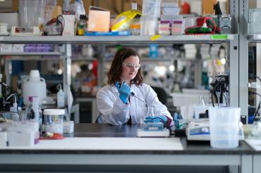
Katheryn Rothenberg, a postdoctoral researcher in U of T's Quantitative Morphogenesis Lab, was lead author on the new study (photo by Qin Dai)
Published: June 7, 2023
Researchers from the University of Toronto's Faculty of Applied Science & Engineering have made progress in understanding the intricate cellular processes involved in tissue development and repair.
The findings, published in the journal Current Biology, shed light on the mechanisms underlying collective cell migration – a fundamental behaviour that plays a crucial role in both normal embryo development and pathological conditions such as cancer metastasis.
“This study advances our understanding of the molecular signals that coordinate cellular behaviours, in embryonic development and tissue repair, and likely also in tumour invasion,” says Rodrigo Fernandez-Gonzalez, a professor in the department of cell and systems biology and the Institute of Biomaterials and Biomedical Engineering who heads the Quantitative Morphogenesis Laboratory.
Researchers found that Rap1 – a molecular switch that regulates cell adhesion and signalling when turned on – plays a role in the formation and remodelling of adherens junctions (protein complexes that occur at cell–cell junctions and cell–matrix junctions in epithelial and endothelial tissue) and the cytoskeleton during the collective cell movements that drive the rapid, scar-less wound healing response in embryos, making it an attractive therapeutic target in the future.
In embryonic wound healing, the cells around the wound move together to seal the lesion. To that end, cells undergo a series of intricate molecular changes. At the centre of these changes, a unique structure called tricellular junction (TCJ) is formed. The TCJ acts as a hub that hosts a series of proteins that are essential in coordinating cell movements.
When researchers tagged the Rap1 protein with a sensor that could be detected by a microscope, they were able to visualize large concentrations of the protein accumulating around the wound, and specifically at the TCJs.
Upon establishing the localization of Rap1 in the hub of wound repair, the researchers set out to find its role in this complex process. By inactivating or reducing the amount of Rap1 in the embryo, they observed a significant reduction in the wound closure rate compared to normal embryos. Conversely, by activating Rap1, the wound closure rate was dramatically accelerated.
“The fact that collective migration speed can be modulated by Rap1 activity provides a potential pathway for either promoting cell migration – for example, to heal chronic wounds or stopping undesired migration like cancer metastasis,” says Katheryn Rothenberg, a postdoctoral researcher in Fernandez-Gonzalez’s lab who led the study.
Researchers also found that Rap1 plays a crucial role in interacting with cell-cell adhesion proteins necessary to maintain cells together as they move to close the wound, and cytoskeletal proteins that cells use to pull on each other and move collectively. They observed that any disruption to Rap1 can greatly impede the speed at which wounds close.
“By unravelling the intricate molecular mechanisms involved, we have uncovered potential targets for therapeutic interventions in various conditions that rely on collective cell migration,” Fernandez-Gonzalez says.
“We are now keen on understanding the upstream signals that turn Rap1 on during wound healing. This understanding would facilitate the development of tools to activate Rap1 in congenital disorders associated with deficient collective cell behaviour, or to inhibit Rap1 when it contributes to spread disease.”


