Not the dress that broke the Internet but still pretty nifty
Published: March 13, 2015
It's the reason television news anchors shouldn't wear striped ties and checked shirts: the moiré effect.
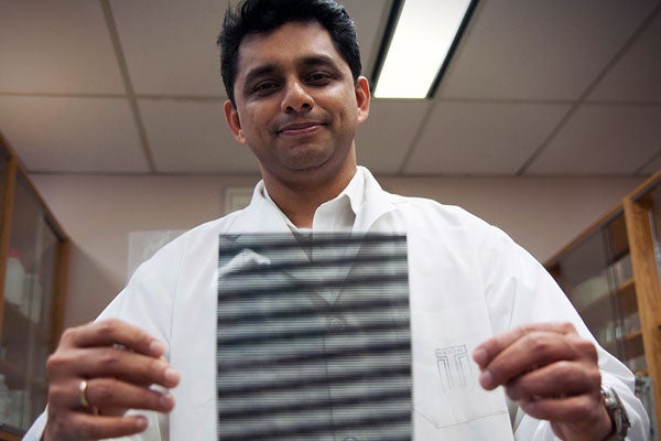
And it's the driving force behind a new laser measurement technique that could help develop non-invasive methods for studying tooth structure, says Dr. Anil Kishen, head of endodontics at the Faculty of Dentistry.
A project first conceived during Kishen’s doctoral work in biomedical engineering at Singapore’s Nanyang Technological University (NTU), this two-laser beam technique offers researchers the ability to study hard tissues under clinically realistic loads on teeth. It also allows them to track the resulting stiffness and breakage within the microstructures of the dentin and enamel (the hard outer shell and inner shell covering the roots of the tooth) – even at the hard-to-measure junction between these two structures – and with pinpoint accuracy.
“Traditionally, to study the effects of force on dental structures you just mount a sample in a machine and apply loads markedly higher than physiological loads,” says Kishen. “But what you end up with is material properties such as stiffness averaged over the entire test specimen.
“This is not accurate, since stiffness differs at every point in the dentin.”
Kishen’s new imaging technique relies upon something called optical interferometry – essentially, a wave-based investigative technique more common to engineering – that results in a whole-field analysis of the tooth and its microstructure with extremely high resolution and under realistic conditions of force, natural for the human mouth.
Enter television’s moiré effect
During experiments, a laser beam splits across the surface of a tooth sample in two parallel lines, which interacts with the hard tissue and creates a virtual grid of the tooth structures at a resolution of 2400 lines per millimetre.
The measurements are transferred to a digital output in dark and light lines known as a moiré pattern. On television, such an effect makes superimposed lines or dots seem to jump and blur. But in dentistry, the result is a complex map representing different levels of strain placed on the microstructure of the tooth. When the lines or “fringes” in the digital output are clumped close together, the strain on the tooth microstructure is significant. When the fringes appear further apart, the strain on the tooth is diminished.
Kishen says this is more than a useful tool for general wear-and-tear studies on teeth; the sensitivity of this imaging technique offers a much greater potential for dentistry research.
“There’s a movement in dentistry towards root canals becoming minimally invasive,” he says, to lessen the risk of tooth fractures after the procedure. “This system can be used to understand how the mechanical properties of the tooth – its toughness – can be improved to prevent root fractures before they occur.”
Kishen’s next step involves using the technique to study the application of non-invasive treatments of infected teeth with nanoparticles.
“We want teeth to be functioning in our patients’ mouths for a long time,” he says.