Neuroanatomy course takes a personal approach to brain illustrations
Published: March 28, 2022
A medical illustrator may well be asked to create illustrations of the brain over the course of his or her career – but rarely do the assignments get personal.
Shelley Wall, associate professor of biomedical communications and biology at the University of Toronto Mississauga, says she wanted to challenge the design and rendering skills of graduate students when the “Neuroanatomy for Visual Communication” course was rescheduled from the master of science in biomedical communications program’s first year to its second year.
The course teaches students the structure and function of the brain and the cranial nerves. They engage in interactive exercises, examine brain specimens and skulls, study historical and contemporary texts, and watch videos of dissections.
"In first year, students are still finding their way in anatomical illustration,” says Wall, adding that the course’s first assignment had previously been a straightforward illustration of the brain in isolation. “Now that Neuro is a second-year graduate course, I could take it to the next level because the students' skills are so much more developed.
“So, I conceived the neuro self-portrait assignment."
Making the assignment a self-portrait not only raised the bar in terms of understanding, it also allowed the students to show off their creativity.
“Drawing the brain is one thing – you must make it accurate,” Wall says. “But what makes this assignment so different is that you really must understand all the important relationships between the brain, the brain case, and the external features of the head. And making the assignment a self-portrait is a way of making it also a completely unique illustration that really puts the students’ stamp on it.”
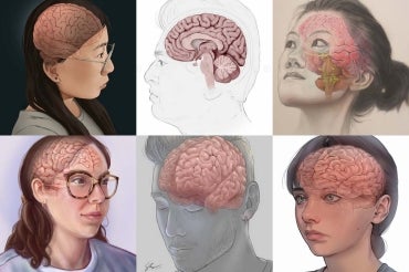
Even working within the constraints of the course assignment, and the strict parameters of depicting the brain with accuracy, the course’s second-year graduate students delivered a broad range of unique and original illustrations.
Mimi (Yuejun) Guo used two different traditional mediums and then digitally composited them to create her self-portrait.
“I used carbon dust to create a black-and-white self-portrait with less saturation and colour to not compete with the brain illustration,” Guo says. “I used acrylic paint for its vibrant colours and to highlight the brain.”
Guo also added a whole new layer of complexity to the assignment by portraying the brain from an upward angle, and at a three-quarter view.
“I chose this perspective to show all the crucial anatomical parts – the cerebral hemisphere, the cerebellum, the brainstem and the origins of the cranial nerves,” says Guo.
One student in Wall’s course worked with brain imaging data belonging to his father, who was diagnosed with a pituitary tumour two years ago and received copies of his MRI scans.
"Before my father's surgery in 2021, I tried to help him understand his condition better,” says Shehryar Saharan. “I was shocked by the lack of high-quality visuals available to explain the tumour in relation to the optic nerve and the rest of the brain. When this neuro assignment was introduced, it became the best excuse to help fill this void and create a neuroanatomy visualisation that would explain my dad's condition in a meaningful and simplified way.”
Saharan asked his father to pose for the portrait and he used his father's brain scans plus many other references to illustrate the brain and the tumour.
Saharan says that his father, whose surgery was a success, was thrilled with his neuro portrait. "After my dad saw the finished piece, he was better able to understand what he had gone through,” Saharan says. “He said he wished that he had had it earlier."
Sana Khan’s brother posed for her portrait, which Khan created in a style that references the 19th-century anatomical atlas Traité complet de l'anatomie de l'homme written by Jean-Baptiste Marc Bourgery, and illustrated by Nicolas Henri Jacob.
“Rather than “ghost” the brain over the portrait, I wanted my illustration to look in vivo – as if you could pull back flaps of skin and tissue to see the brain within,” says Khan.
The homage to Bourgery’s canonical text also adds a touch of whimsy to the illustration, Wall says. She adds that, for medical illustrators, the neuro self-portrait assignment, which can be viewed on the program’s Instagram account, is the perfect intersection of complexity and accuracy, and creativity and originality.
“I like to think that this assignment not only challenges the students, but let’s them have some artistic fun as well.”


