Fighting cancer: U of T researchers develop new tool to track circulating tumour cells
Published: November 21, 2016
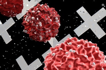
Cancerous tumours are known to release cells into the bloodstream, and it is these circulating tumour cells (CTC) that are the sources of metastatic tumours – tumours that spread and form in distant locations in the body and can eventually kill patients.
A breakthrough by Professor Shana Kelley’s research group at the University of Toronto published in Nature Nanotechnology provides a new tool to characterize CTCs that may help cancer biologists and clinicians understand how to use these cells to provide better treatment.
Monitoring circulating tumour cells has been a tremendous challenge as they are outnumbered in blood by healthy cells at a level of over 1 billion-1. Moreover, they can display varied and dynamic properties, and the collection of CTCs found in the bloodstream of a cancer patient may have differing metastatic potential. Consequently, efforts to integrate the analysis of these cells into mainstream clinical medicine have been limited because it has been difficult to pinpoint what types of cells and what phenotypic properties should be targeted.
But the potential of CTCs to allow the collection of a non-invasive “liquid biopsy” to monitor cancer progression is a tantalizing possibility that has continued to attract significant attention to this problem.
The Kelley research group found that by using magnetic nanoparticles, CTCs in blood samples could be targeted based on proteins displayed on the cell surface, and separated based on the levels of the protein present. Using a high–resolution microfluidic device, cells were then separated into 100 different capture zones to generate a profile that provides phenotypic information related to metastatic potential.
Using this approach and monitoring cells generated in animal models of cancer and in samples collected from prostate cancer patients, the properties of CTCs were shown to evolve and become more aggressive as tumours became more advanced.
“Through this approach, we aimed to provide a new way to profile CTCs beyond simply counting their numbers in clinical samples,” explained Mahla Poudineh, lead author of the paper who is a graduate student at the Faculty of Applied Science & Engineering. “Instead, we wanted to provide phentotypic information that might allow these cells to be classified as benign or more dangerous, which would then inform treatment options.”
Kelley is a professor at the Faculty of Pharmacy and the Institute for Biomaterials and Biomedical Engineering. The Kelley group along with collaborators at the Sargent Group, Faculty of Applied Science & Engineering Professor Ted Sargent's research lab, hope to turn the approach they reported into a device that can be used by cancer researchers and eventually clinicians to allow CTC analysis to be monitored routinely and used to limit the progression of cancer.
“We were very fortunate to collaborate with a number of oncologists at the Sunnybrook Research Centre and Princess Margaret Hospital as we developed this technology so that we could test our approach with real patient specimens and better understand how to adapt it for use in the clinic,” Kelley said.